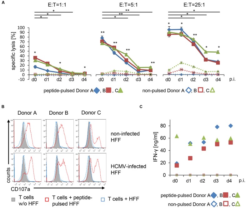
FIGURE 3.
T cell receptor-mediated lysis of HFF is reduced by HCMV starting from 1 day after infection. (A) TCR-mediated lysis of non-infected and HCMV-infected HFF (AD169 ΔUS2-11, MOI 5) by EBV-specific T cells (E:T = 1:1; 5:1; 25:1) was analyzed in a 4 h Eu release assay at the indicated time points after infection (p.i. = after infection; three technical replicates with each donor). Target cells were pulsed with BMLF1-peptide (2 μM GLCTLVAML) for 2 h before co-incubation with EBV-specific T cells or left untreated. (B) The histograms show the degranulation of EBV-specific T cells after co-incubation with either untreated or peptide-pulsed non-infected and HCMV-infected HFF (AD169 ΔUS2-11, MOI 5, d4 p.i.), as analyzed by flow cytometric detection of CD107a at the cell surface. (C) Levels of IFN-γ secreted into the supernatants of co-cultures of EBV-specific T cells and peptide-pulsed or non-pulsed HFF, respectively (non-infected or 1–4 days after infection with AD169 ΔUS2-11, MOI 5, as indicated).

