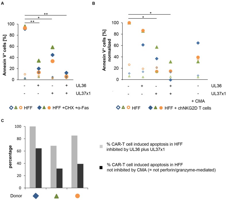
FIGURE 5.
UL36 and UL37x1 inhibit CAR-T cell-mediated apoptosis in HFF. The anti-apoptotic HCMV-encoded proteins UL36 and UL37x1 were expressed in HFF by either mRNA electroporation or retroviral transduction, respectively. (A) The anti-apoptotic function of the exogenously expressed proteins was confirmed by overnight incubation of the HFF with cycloheximide (CHX, 10 μg/ml) plus an agonistic anti-Fas-antibody (0.2 μg/ml). A GFP encoding vector or mock-electroporation were used as controls. The diagram shows the fraction of apoptotic HFF determined by Annexin V staining in a flow cytometric analysis (n = 3). (B) The diagram shows the induction of cell death in HFF expressing UL36 and/or UL37x1 after 4 h of co-incubation with chNKG2D-expressing αCD3/αCD28-activated T cells (n = 3, 3 different T cell donors). The percentages of apoptotic HFF obtained in the co-cultures of CAR-T cells plus HFF without UL36/UL37x1 expression were set to 100%. Concanamycin A (CMA, 100 nM) was used to block perforin-induced apoptosis. (C) The diagram displays the percentage of CAR-T cell-mediated apoptosis in HFF that was inhibited by the combined expression of UL36 and UL37x1 (gray bars; obtained by subtraction of the respective values shown in (B) from 100%). The black bars show the proportion of CAR-T cell induced apoptosis in HFF, which was blocked by CMA (B) i.e., the cell death that was not mediated by perforin/granzyme. If these values were a result of incomplete CMA blockade, then the black bars (non-perforin/granzyme mediated cell death) would be even lower. The respective differences between the gray and the black bars correspond to the proportion of the perforin/granzyme-mediated cell death in the HFF, which was inhibited by the combined expression of UL36 and UL37x1.

