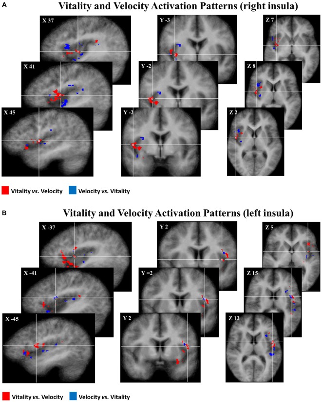
Figure 5.
Maps group of 50% of most discriminative voxels for the perceptual difference of vitality forms (red) and velocity (blue) collapsing three different contrasts (rude vs. fast, neutral vs. medium, gentle vs. slow) in the right (A) and in the left (B) insula. Each voxel was reported if it was present in at least 10 of the 16 participants. These activation patterns (pFDR < 0.05) are overlaid on the average anatomical template of 16 participants in Tailarach coordinates.

