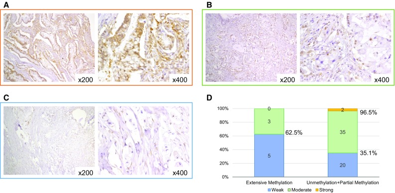
Fig. 3.

Expression analysis of EFEMP1. IHC staining of EFEMP1 in intraductal papillary mucinous neoplasms with strong staining (a), moderate staining (b), and weak staining (c). Association between EFEMP1 methylation status and IHC staining (d). EFEMP1, epidermal growth factor-containing fibulin-like extracellular matrix protein 1; IHC immunohistochemical; NA not available
