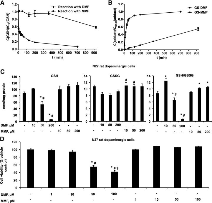
Figure 1.
DMF forms an adduct with GSH and depletes intracellular GSH levels and decreases cell viability in a dose-dependent manner. Time course of the S-alkylation reaction between 1 mm DMF or 1 mm MMF with 1 mm GSH in PBS at pH 7.4 was measured as described in Materials and Methods. Time course for GSH consumption (A) and GS-DMF and GS-MMF adduct formation (B). C, Intracellular GSH, GSSG, and their ratio (GSH/GSSG) were determined at 24 h after DMF and MMF (10, 50, and 200 μm) treatment. Bars represent mean ± SEM. *p < 0.05 compared with controls; #p < 0.05 compared with DMF at 10 μm (n = 4). D, Cell viability in N27 rat dopaminergic cells treated with DMF (1, 10, 50, and 100 μm) or MMF (1, 10, 50, and 100 μm) for 24 h was assessed using Presto Blue cell viability kits. Bar plot represents the percentage of the control as mean ± SEM of viable cells (n = 3). *p < 0.05 compared with control (DMSO); #p < 0.05 compared with the DMF (1 μm)-treated group; $p < 0.05 compared with the DMF (50 μm)-treated group (n = 3).

