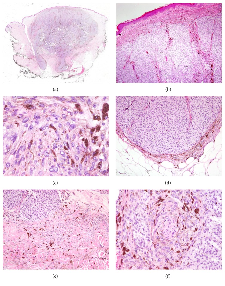
Figure 1.
A pigmented lesion consisted with cellular blue nevus with an expansive pattern of extension into dermis and subcutaneous adipose tissue (a) and a spared papillary dermis (b). Cellular islands of closely aggregated spindle shaped cells with ovoid nuclei revealed a mild nuclear enlargement, a mild pleomorphism, and prominent small nucleoli (c). The deepest boundary of the tumor was a pushing border (d) with foci of more irregular infiltrative borders (e). Perineural invasion was observed (f).

