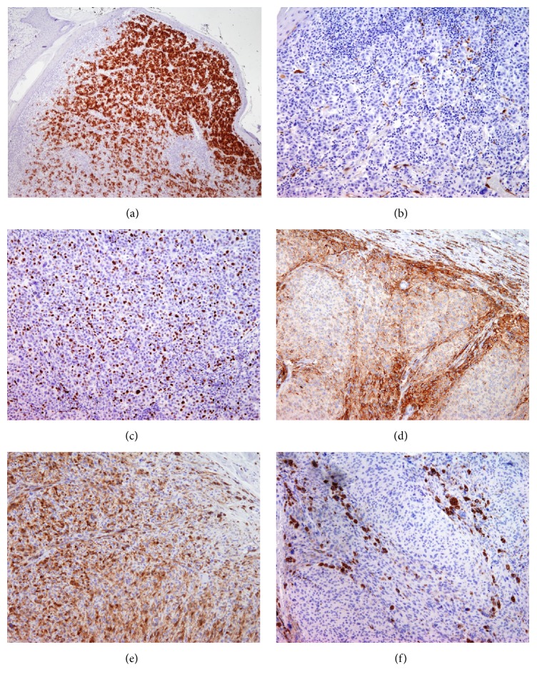
Figure 3.
Immunohistochemical staining of nodular melanoma (NM) and cellular blue nevus (CBN) with HMB-45, p16, and Ki-67: HMB-45 was strongly positive in NM (a) while in CBN the reactivity was mostly present at the periphery of alveolar nets (d); p16 was negative in NM (b) but present in the majority of tumor cells in CBN (e); Ki-67 was positive in 33% of melanoma cells (c) and negative in CBN (f).

