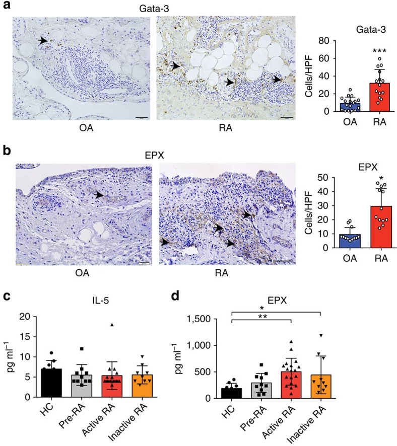
Figure 6. Expression of Th2 and eosinophil markers in human rheumatoid arthritis.
(a) Representative immunohistochemistry staining of GATA3 in the synovium of osteoarthritis (OA) and RA patients. Positive cells per high-power field were compared between groups (n=17 OA patients and 14 RA patients). (b) Representative immunohistochemistry staining of EPX in the synovium of OA and RA patients. Positive cells per high-power field were compared between groups (n=12 OA patients and 12 RA patients). (c) IL-5 serum levels in healthy controls, autoantibody-positive individuals without RA, active and inactive RA patients (n>10 patients per group). (d) Serum EPX levels in healthy controls, autoantibody-positive individuals without RA, active and inactive RA patients (n>10 patients per group). (*P<0.05, **P<0.01 and ***P<0.001 determined by Student's t-test for single comparison (a,b) or analysis of variance test for multiple comparisons (c,d)).

