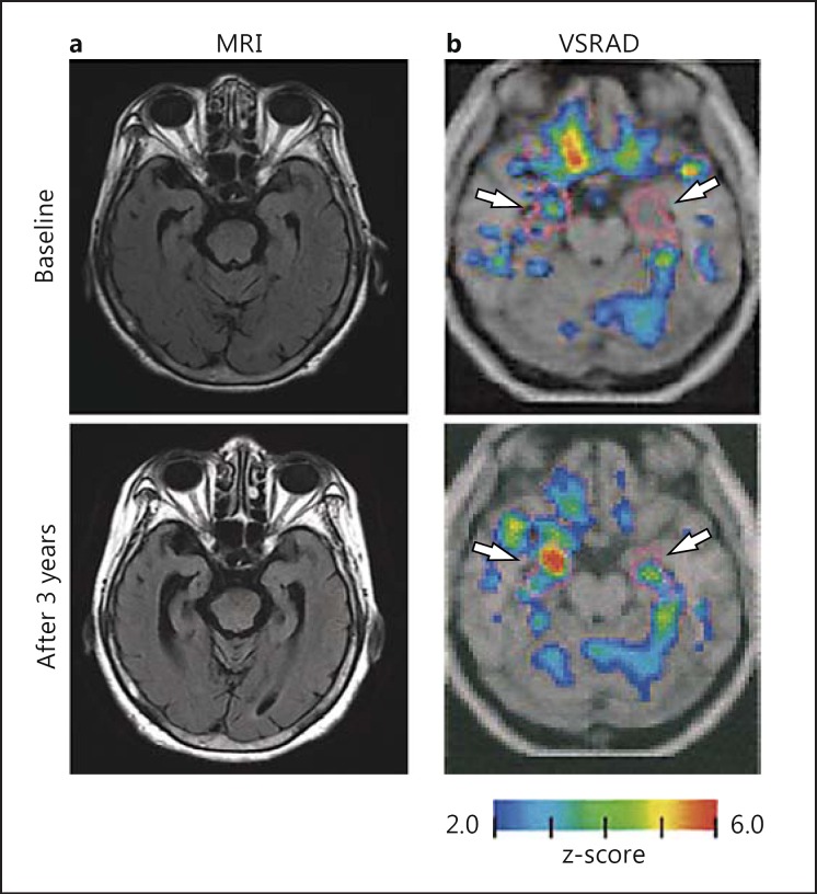
Fig. 1.
Changes in severity of gray matter atrophy in the VOI (HAI) during the 3-year follow-up in the case of a 78-year-old female. a MRI. Progression of medial temporal lobe atrophy was observed on FLAIR MRI during the 3 years. b VSRAD. Colored areas (colors refer to the online version only) with z-scores of >2 are overlaid as significantly atrophied regions on the standardized MRI template. The target VOI is surrounded by purple lines (white arrows), and red areas indicate higher gray matter atrophy levels as compared with the control (normal database). The HAI (z-score) increased from 1.57 to 3.20, and the MMSE dropped from 23 to 17 after 3 years.

