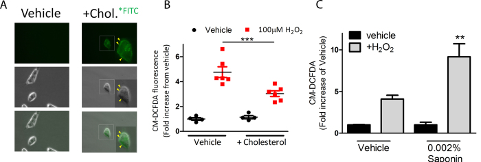
Figure 3. Altering the membrane permeability of breast cancer cells using light detergent or cholesterol affects uptake of ROS.
(A) Representative bright field and fluorescent microscopy shows MDA-MB-231 cells treated with cholesterol tagged with fluorescein or vehicle control for 2 hours before washing off excess. Fluorescent microscopy indicates localization of cholesterol in the membrane of cells (yellow arrows). (B) Quantification of flow cytometric data shows % increase of intracellular ROS following incubation with cholesterol (2 h) and 100 μM H2O2 (30 min) as determined by activity of the fluorescent probe, CM-DCFDA. (C) Quantification of flow cytometric data shows % increase of intracellular ROS following incubation with 0.002% saponin (2 h) and 100 μM H2O2 (30 min) as determined by activity of the fluorescent probe, CM-DCFDA.

