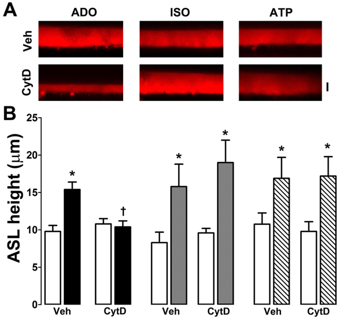
Figure 4. Cytochalasin D abolishes adenosine- but not isoproterenol-mediated ASL secretion in normal HBECs.

HBECs were exposed to vehicle (0.1% DMSO) or 10 μM cytochalasin D (CytD) for 30 min before agonist addition. (A) Typical XZ confocal micrographs of ASL (red) taken after 10 min exposure to adenosine, isoproterenol and ATP (all at 100 μM) in the presence and absence of 10 μM cytochalasin D in normal HBECs. (B) Mean ASL height data. Open bars, control. Closed bars, adenosine; grey bars, isoproterenol; hatched bars, ATP. All n = 6. *p < 0.05 different from vehicle. †p < 0.05 different ± cytochalasin D.
