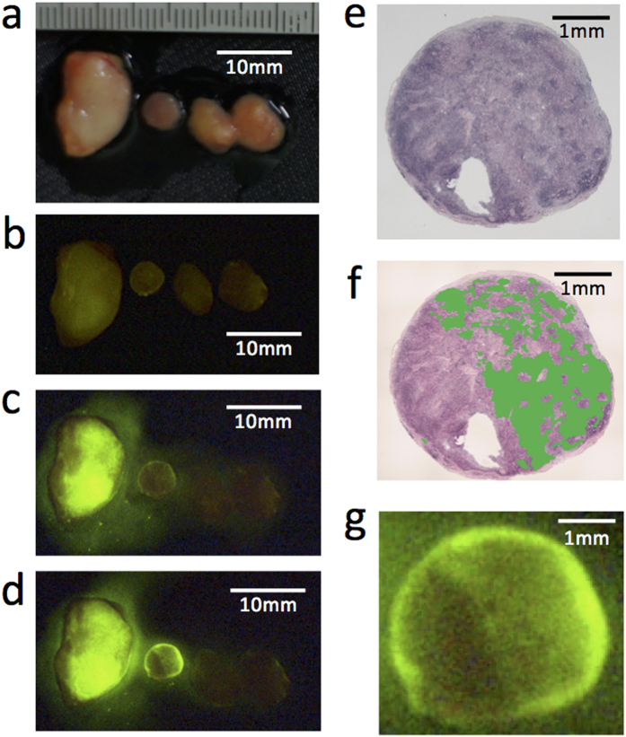
Figure 1. Detection of cancer cells in lymph nodes using gGlu-HMRG fluorescence.
(a) Macroscopic image of resected lymph nodes from one breast cancer surgery case. (b) Fluorescent image of the same lymph nodes as in (a) before administration of gGlu-HMRG. Autofluorescence is indicated by the faint green color. Fluorescent images (c) 5 minutes and (d) 15 minutes after administration of gGlu-HMRG. (e,f) H&E staining of the same lymph node second from the left in (a–d)). This lymph node was diagnosed pathologically as metastatic. (f) Metastatic regions were indicated by green color. (g) Magnified fluorescent image of the same lymph node in (e,f) 15 minutes after administration of gGlu-HMRG.

