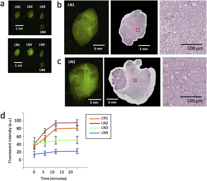
Figure 2. Detection of metastasis in lymph nodes using gGlu-HMRG fluorescence.
(a) Fluorescent image of four resected lymph nodes before (upper picture) and 15 minutes after (bottom picture) administration of gGlu-HMRG. (b,c) Fluorescent images 15 minutes after administration of gGlu-HMRG (left) and H&E staining (middle) of two metastatic lymph nodes. The small yellow circles on the left indicate the ROIs that showed the strongest increase in fluorescent intensity within 5 minutes. The small red boxes in the middle images correspond with the yellow circles on the left. Magnified H&E staining of the regions within the red boxes are shown on the right, and these were diagnosed as cancerous lesions by pathological examination. (d) Time-dependent increases in the fluorescent intensity of each lymph node after administration of gGlu-HMRG.

