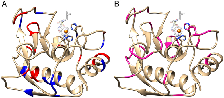
Figure 2.
(A) The X-ray structure of catMMP12 (1RMZ42, which does not carry the R5 fusion) showing the charged residues in blue (positive) and red (negative). (B) The structure of catMMP12 with the residues showing no signals shown in magenta. Apparently, they tend to cluster close to loops or in proximity of charged residues, which can interact with the biosilica matrix.

