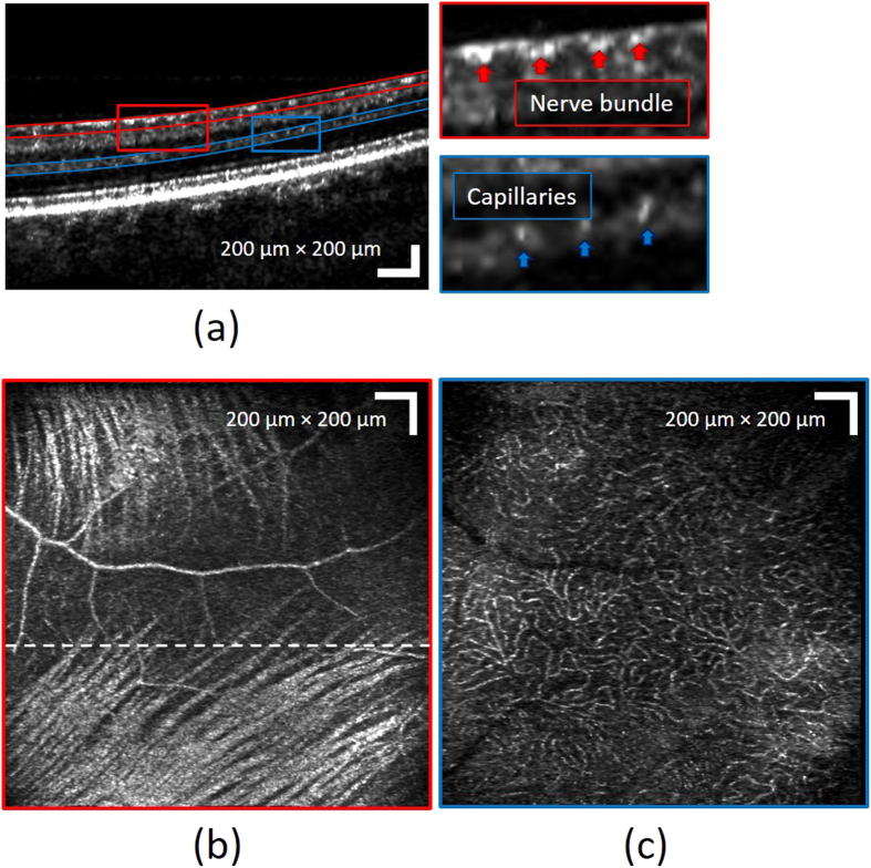
Figure 7. Images of different retinal layers acquired with the MAL-WSAO-SS-OCT.
(a) The wide-field B-scan image is presented on a logarithmic scale. The red and blue curved lines indicate the depths used for generation of the en face images, (b) the retinal nerve fiber layer and (c) laminar capillary bed, respectively.

