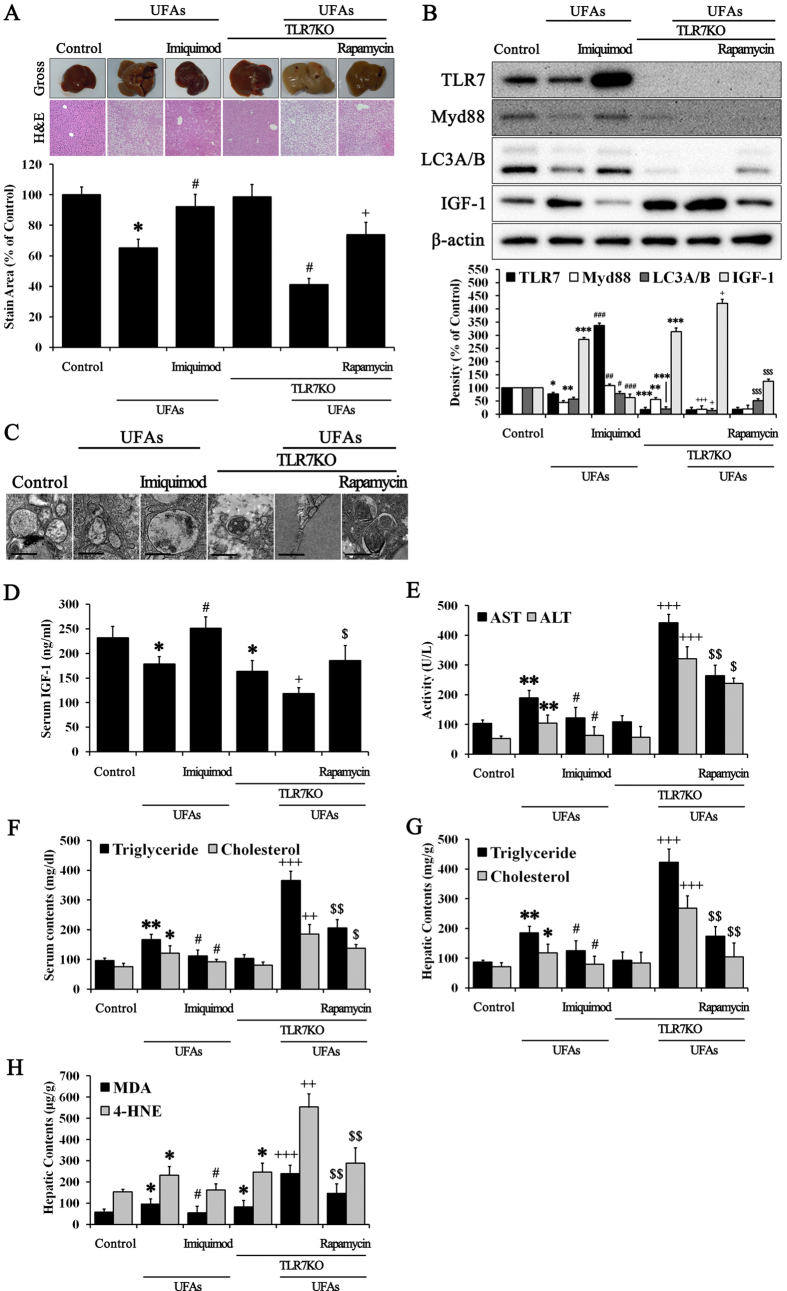
Figure 5. NAFLD was improved by TLR7-mediated regulation of IGF-1.
Normal control mice and TLR7KO mice were injected with or without 0.1 mg/kg imiquimod and 2 mg/kg rapamycin twice a week while eating either a UFAs diet or a normal research diet for 8 weeks. (A) Histopathology analyses were performed, and we photographed the gross appearance of the liver and tissue section images stained with H & E (Indicated magnification: X200). The stained area was quantified with the Image Pro analysis program. Immunoblotting was performed to evaluate (B) the protein expression of TLR7, Myd88, LC3A/B, IGF-1, and β-actin in liver tissues. (C) Images of autophagosome formation were obtained from TEM in corresponding groups. The scale bar indicates 500 nm. Serum contents of (D) IGF-1, (E) AST, ALT, (F) triglycerides, and cholesterol were examined. Hepatic contents of (G) triglycerides, cholesterol, (H) MDA, and 4-HNE were examined. Data are mean and SEM values (n = 3). *p < 0.05 vs. untreated control mice. **p < 0.01 vs. untreated control mice. ***p < 0.001 vs. untreated control mice. #p < 0.05 vs. control + UFAs group. ##p < 0.01 vs. control + UFAs group. ###p < 0.001 vs. control + UFAs group. +p < 0.05 vs. TLR7 group. ++p < 0.01 vs. TLR7 group. +++p < 0.001 vs. TLR7 group. $p < 0.05 vs. TLR7KO + UFAs group. $$p < 0.01 vs. TLR7KO + UFAs group. $$$p < 0.001 vs. TLR7KO + UFAs group.

