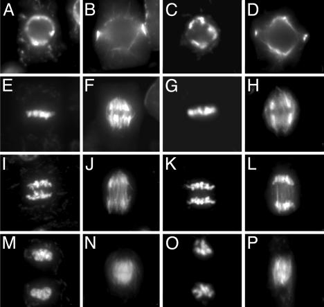
Fig. 1.
Mitosis in root tips of wild-type (A, B, E, F, I, J, M, and N) and atrad51-1 (C, D, G, H, K, L, O, and P) seedlings. Chromosomes (Left) and microtubule structures (Right) in the same cells were visualized with 4′,6-diamidino-2-phenylindole, and antibodies against β-tubulin were visualized at different stages during the mitotic cell cycle (preprophase, A and C; metaphase, E and G; anaphase, I and K; and telophase, M and O).

