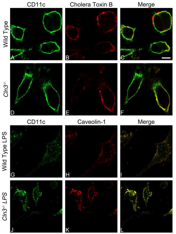
Fig. 6. Localization of CD11c to lipid rafts in unstimulated or LPS-stimulated Cln3−/− and wild type bone marrow cultured cells assessed by fluorescence-based immunocytochemistry.
Cln3−/− and wild type bone marrow cultured cells (BMCCs) were assessed for surface CD11c and lipid raft markers with fluorescence-based immunocytochemistry without membrane permeabilization, and imaged with confocal microscopy using a 100× objective lens. Representative images are shown. Unstimulated mixed BMCCs were labeled with an anti-CD11c antibody (green fluorescence, A, D) and lipid raft binding cholera toxin B (CTB; red fluorescence, B, E). LPS-stimulated BMCCs were labeled with antibodies against CD11c shown in green (G, J) and lipid raft protein caveolin-1 shown in red (H, K). Yellow in the merged images indicates colocalization. Scale bar = 10 microns.

