
Figure 2.
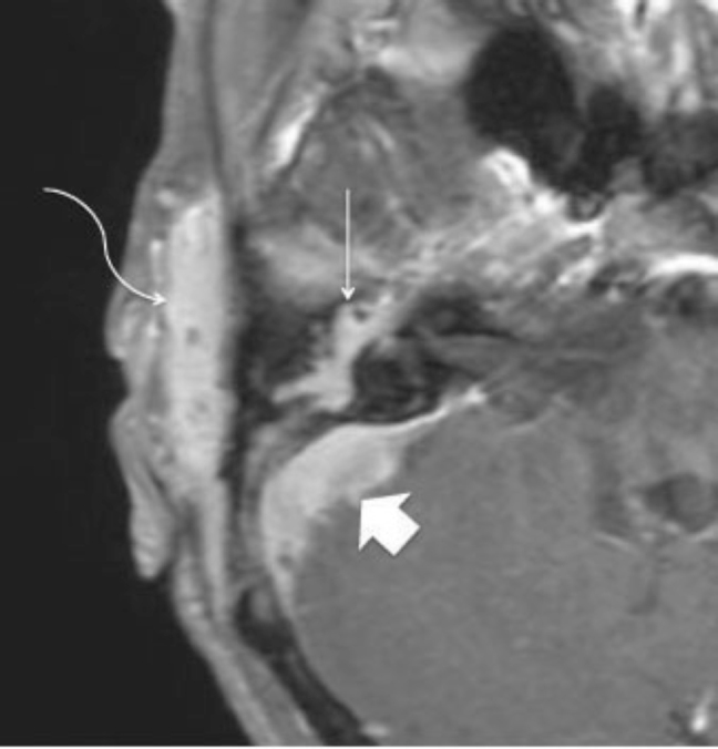
72-year-old woman with clear-cell meningioma. Axial T1 post-Gadolinium MRI. Lateral arrow indicates the enhancing soft-tissue mass involving the peri-auricular subcutaneous tissues. Note peri-auricular soft-tissue infiltration (curved arrow), middle ear involvement incorporating the facial nerve (straight arrow), and intracranial extension involving the sigmoid sinus (arrowhead).