Figure 1.

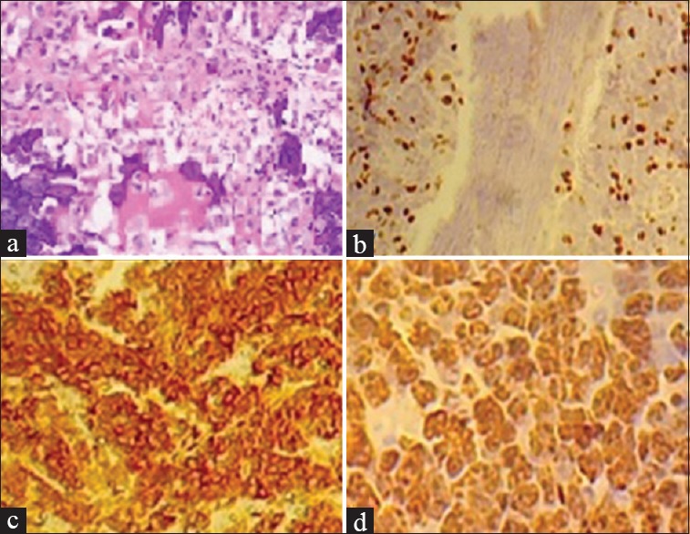
HER2, P53, and Ki67 overexpression in osteosarcoma. (a) Photomicrograph of osteosarcoma (hematoxylin and eosin). Immunohistochemical analysis revealed (b) Ki67 with 30% positivity, and (c) HER2 grade 3+ reactivity. (d) P53 demonstrating clusters of nuclei positivity of neoplastic cells
