Fig. 1.

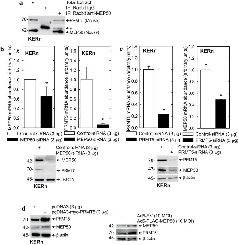
MEP50 and PRMT5 form a complex in keratinocytes. A Freshly isolated foreskin keratinocyte lysates (300 μg) were used for immunoprecipitation with Rabbit IgG or rabbit anti-MEP50, and 10 μg of total extract was electrophoresed. The antibodies for immunoblot are mouse anti-MEP50 and goat anti-PRMT5. Similar results were obtained in three separate experiments. The upper band (*) in the blot probed with MEP50 is non-specific. B/C Keratinocytes were electroporated with 3 μg of control-, MEP50- or PRMT5-siRNA. After 48h, RNA was isolated and MEP50 and PRMT5 mRNA levels were assessed by qRT-PCR. The values are mean ± SEM, n = 3. The asterisks indicate significant differences as determined by the students t-test (*, p<0.005). Extracts were also prepared to assess PRMT5 and MEP50 protein level. D MEP50 or PRMT5 overexpression. KERn were electroporated with 3 μg of control plasmid or plasmids encoding MEP50 or PRMT5, or MEP50-encoding adenovirus (10 MOI). After 48 h protein lysates were prepared for immunoblot with anti-MEP50 and anti-PRMT5. β-actin was used as the loading control. Similar results were observed in each of three experiments.
