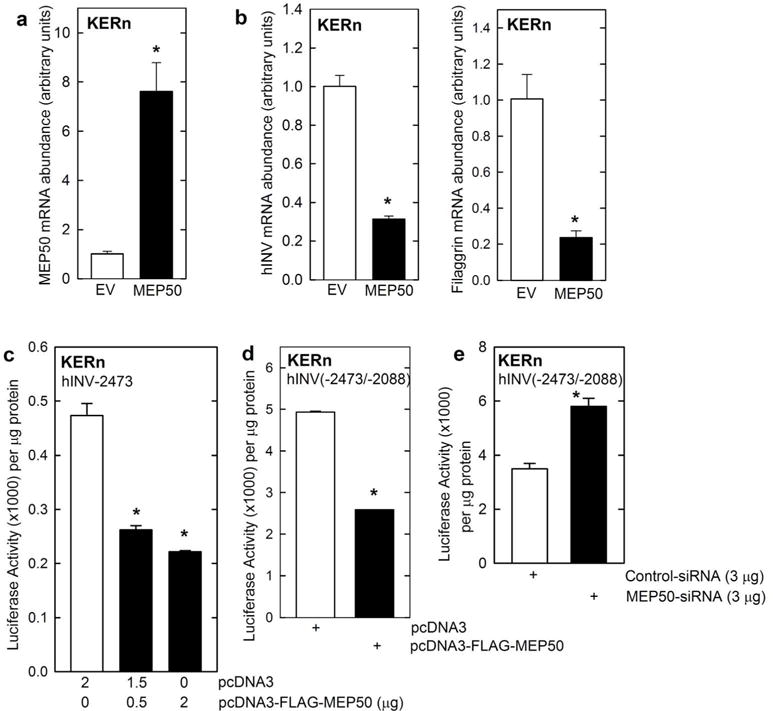
Fig. 2.

MEP50 suppresses involucrin expression. A/B KERn were electroporated with the indicated plasmids. After 24 h RNA was isolated and MEP50, involucrin and filaggrin mRNA levels were assessed by qRT-PCR. The values are mean ± SEM (n = 3). The asterisks indicate a significant difference (p < 0.005). C/D KERn were transfected with 0.5 μg of the indicated involucrin promoter plasmids in the presence of 1 μg of pcDNA3 or pcDNA3-FLAG-MEP50. At 24 h post-transfection, extracts were prepared and assayed for promoter activity. E KERn were electroporated with 3 μg of control-siRNA or MEP50-siRNA. After 48 h, the cells were re-electroporated with 3 μg of endo-free involucrin promoter. After an additional 24 h, extracts were prepared and promoter activity (luciferase) assay.
