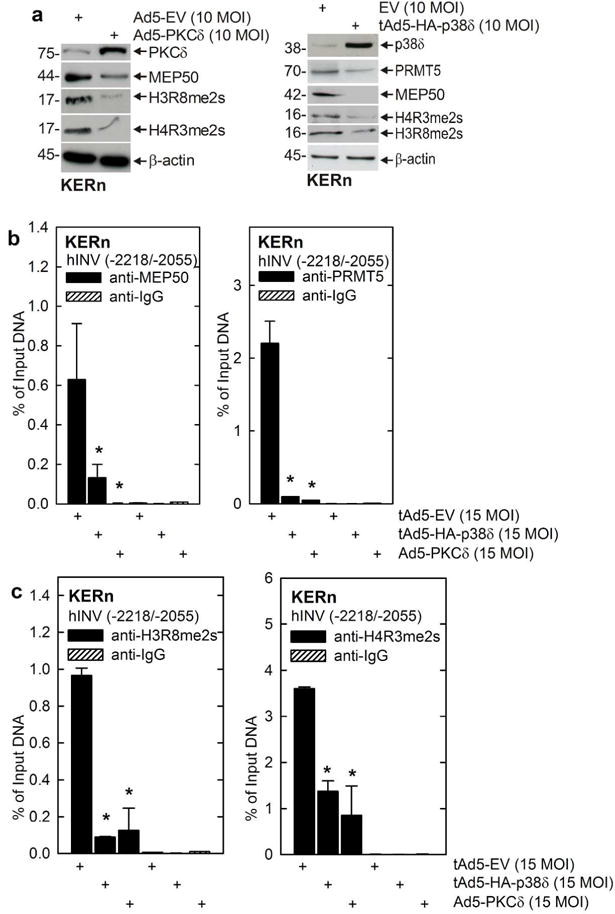
Fig. 5.

PKCδ/p38δ signaling reduces MEP50 and PRMT5 level and hINV promoter activity. A KERn were infected with 10 MOI of tAd5-EV or Ad5-PKCδ and at 48 h extracts were prepared for detection of PKCδ, MEP50, H3R8me2S and H4R3me2s. β-actin was used as a loading control. Similar results were obtained in three different experiments. B KERn were infected as above and after 48 h ChIP was performed using the Diagenode Low Cell ChIP Kit and primers spanning nucleotides -2218/-2055 if the hINV promoter region which includes the AP1-5 site. The values are mean ± SEM, n = 3. The asterisks indicate significant difference (p < 0.005).
