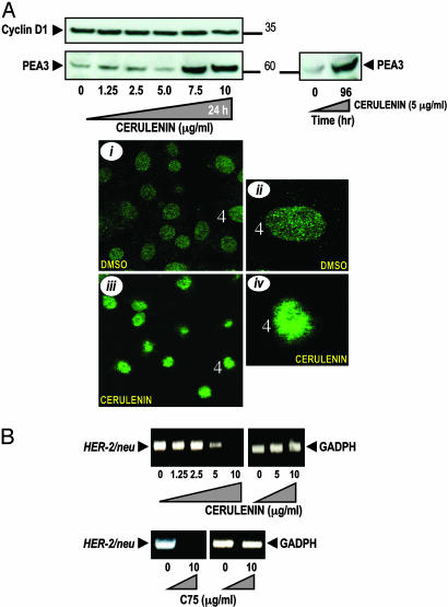
Fig. 2.
Accumulation of PEA3 and inhibition of HER2 gene transcription after pharmacological blockade of FAS activity. (A)(Upper) SK-Br3 cells were cultured either with graded concentrations of cerulenin for 48 h (Left) or exposed to 5 μg/ml cerulenin for 96 h (Right). Fifty micrograms of protein was subjected to Western blot analyses with anti-PEA3 and Cyclin D1 Abs. (Lower) SK-Br3 cells were treated with 2.5 μg/ml cerulenin for 48 h, and subcellular localization of immunofluorescent PEA3 was assessed with a Leica DMIRE2 confocal microscope. PEA3-stained cell nuclei are presented at two magnifications (control, i and ii; and cerulenin-treated cells, iii and iv). (B) Total RNA from cerulenin- and C75-treated SK-Br3 cells was isolated and RT-PCR analyses for HER2 and GADPH transcripts and expression were performed as described in Materials and Methods.

