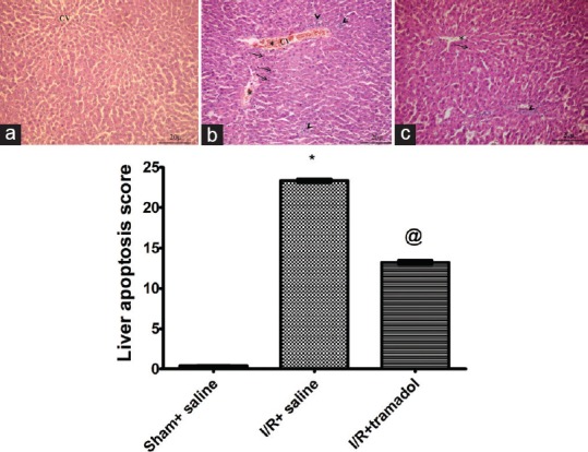
Figure 1.

Photomicrographs are representative of cross sections from six rats stained with hematoxylin and eosin. The sham group (a) showed normal hepatocytes arranged in branching plates radiating from the central vein. The nuclei were central and rounded and the cytoplasm was granular and acidophilic. Normal sinusoidal space and periportal area were also observed. No inflammatory activity could be seen. Ischemia/reperfusion operated rats (b) presented marked congestion (asterix) and cellular infiltrates (arrow heads). Treatment with tramadol in group (c) showed less congestion, hepatocytes-loss, and cellular infiltrates. Bar graph showing liver apoptosis scores, (n = 10) *P < 0.05 (significantly different from Sham group); @P < 0.05 (significantly different from Ischemia/reperfusion group) (a-c) ×20
