
Fig. 1. SRS microscopy workflow and imaging of normal gray and white matter.
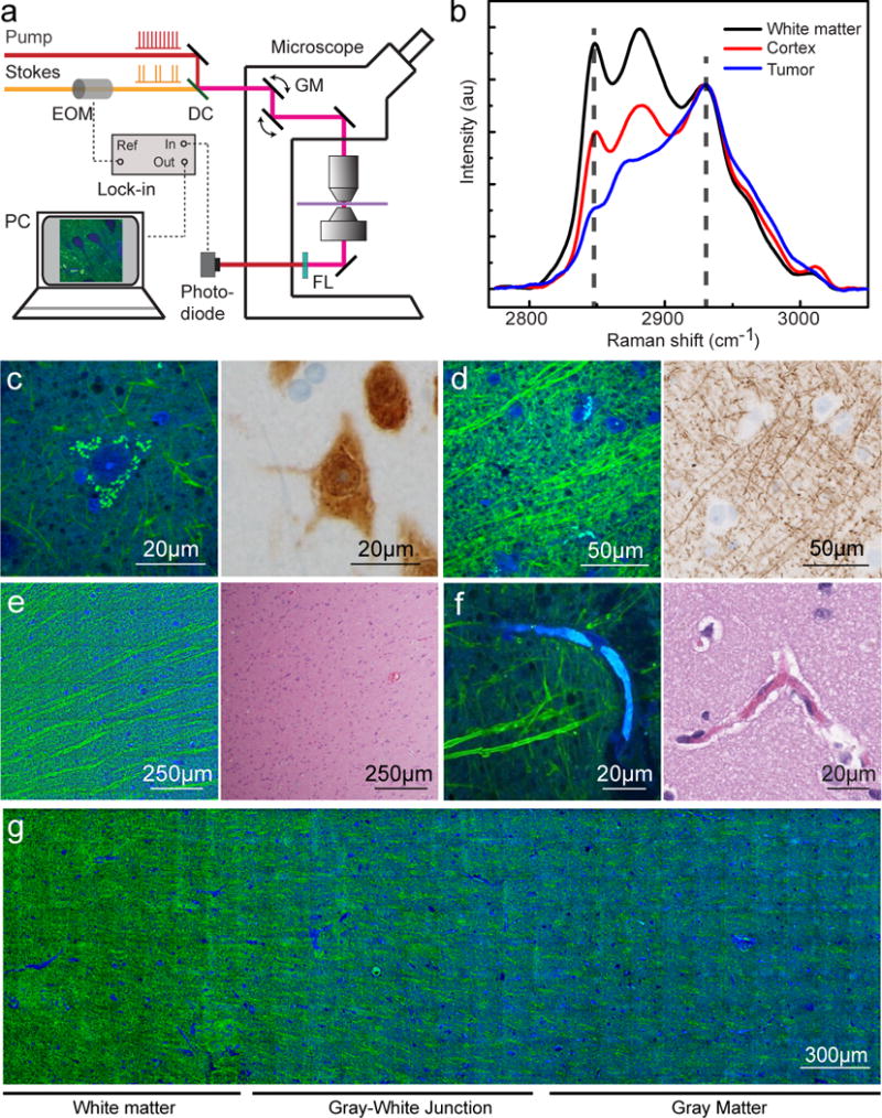
All imaged specimens were collected from patients undergoing anterior temporal lobectomy for intractable epilepsy. (A) Experimental setup of SRS microscopy. The Stokes beam was modulated at high frequency (10 MHz), and the weak stimulated Raman loss signal was demodulated by a lock-in amplifier. A transmission mode detection scheme was used for ex vivo imaging on fresh tissues. DC, dichroic mirror; EOM, electro-optical modulator; FL, optical filter; GM, galvanometer mirror. (B) Raman spectra from fresh sections of human glioblastoma biopsy show white matter, cortex, and tumor. The marked frequencies (dashed lines) at 2845 and 2930 cm−1 were chosen for two-color SRS imaging. (C) SRS imaging of normal gray matter at high magnification showing neuronal soma with pyramidal architecture filled with lipofuscin-rich granules (left), that stain positively for the neuronal nuclei antigen (NeuN) within the neuronal cell body (right). (D) SRS imaging of white matter (left) demonstrates individual axons appearing as linear, lipid-rich structures that correspond well with neurofilament immunohistochemical staining (right). (E) An SRS image of the gray-white junction (left) demonstrates parallel bundles of lipid-rich white matter tracts that are not visible with H&E staining (right). (F) Capillaries filled with protein-rich erythrocytes appear blue on SRS imaging (left) and eosinophilic on H&E-stained section (right). (G) At low magnification, the biochemical differences between protein-rich gray matter (blue) and myelinated white matter (green) are apparent.