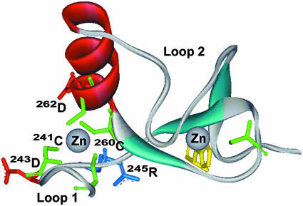
Fig. 5.
Model of S-nitrosylation of parkin. Ribbon structure of a partial sequence of human parkin showing RING I domain (residues 237Thr to 291Ala). α-Helix shown in red, β-sheet in cyan, cysteine residues in green, histidine in yellow, aspartate in red, and arginine in blue. 241Cys and 260Cys match the nitrosylation consensus motif (13).

