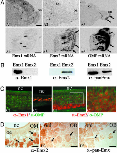
Fig. 1.
Emx2 is expressed in the adult mouse OSNs. (A) In situ hybridization of Emx1 (A1 and A4), Emx2 (A2 and A5), and OMP (A3 and A6) mRNAs by using 35S-labeled oligonucleotide probes and film autoradiography (10 days for Emx1 and Emx2 and 4 days for OMP) was performed on brain (A1–A3) and olfactory mucosa (A4–A6) cryostat sections. Emx1 mRNA is detected in the olfactory bulb (OB) and cortex (Cx) but not in the olfactory mucosa (A4). Emx2 mRNA is not detected in the olfactory bulb or cortex (A2) but is visualized in the olfactory mucosa (A5, arrows) with a pattern similar to that of OMP mRNA (A6, arrows). As reported in refs. 26 and 44, OMP mRNA is also detected in the olfactory nerve and axon terminals in the olfactory bulb (A3, arrows). (Scale bar, 1 mm.) (B) Characterization of the anti-Emx1, anti-Emx2, and pan-Emx antibodies on extracts of COS-7 cells overexpressing Emx1 or Emx2. Anti-Emx1 and anti-Emx2 antibodies detect Emx1 and Emx2, respectively, whereas pan-Emx antibody recognizes Emx1 and Emx2. (C) Colocalization of Emx1 or Emx2 (in red) with OMP (in green) in olfactory mucosa cryostat sections fixed with low paraformaldehyde. The Emx1-specific antibody labels the nucleus of small cell clusters observed mainly in thin areas of the olfactory epithelium (C2) and very rarely in the principal pluristratified epithelium containing most OSNs (C1). The Emx2-specific antibody labels the nucleus of virtually all OSN (C3). Emx2 is found in the nuclei of immature and mature neurons. Immature OMP-negative neurons in the basal part of the epithelium display a higher nuclear labeling (C3) than the mature OMP-positive neurons in the medial and apical part of the epithelium (C4, which is the area indicated by a square in C3). nc, nasal cavity. (Scale bar, 20 μm.) (D) Immunoperoxidase detection of Emx2 in olfactory mucosa (OM) and bulb (OB). Anti-Emx2 antibody labels the axonal tracts (D1, arrows) and glomeruli (D2, arrows). The same localization was obtained with the pan-Emx antibody (D3). oe, olfactory epithelium; onl, olfactory nerve layer. (Scale bar, 80 μm.)

