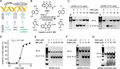
FIGURE 1.
Rhein selectively inhibits AlkB in vitro. A, the three major repair pathways in E. coli. DNA glycosylase is colored in cyan, DNA methyltransferase repair is in blue, and the AlkB repair is in green, respectively. B, scheme of AlkB repair methylated DNA. The structures of the inhibitor rhein and negative control BA are shown. C, rhein inhibits AlkB repair of m1A in ssDNA (left panel) and dsDNA (right panel) by using the DpnII digestion assay. The upper band is 49-nucleotide dsDNA, which contains an m1A lesion, and the lower band represents the mixture of 22- and 27-nucleotide dsDNA products after DpnII digestion. The 2OG concentration is 50 μm. D, quantitative determination of inhibitory activity using HPLC-based assay. The fitted IC50 is 12.7 μm assayed at 50 μm 2OG. The error bars are the means ± S.E. (n = 3). E, BA fails to inhibit m1A-dsDNA repair by AlkB. The 39-nucleotide m1A-containing DNA substrate was tested. F, C-Ada repair of O6mG is unimpaired in the presence of rhein. The upper band is 39-nucleotide O6mG-containg dsDNA, and the lower band is the digested fragments by PvuII. G, rhein is inactive to inhibit AlkA glycosylase. The 25-nucleotide mismatched dsDNA substrate is tested. All reactions were assayed in triplicate. nt, nucleotide.

