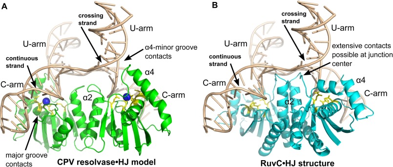
FIGURE 7.
Comparison of a CPV resolvase·HJ complex model with the RuvC·HJ complex. A, model of a CPV resolvase·HJ complex obtained by superposition of the resolvase dimer onto the RuvC dimer shown in B. B, structure of the T. thermophilus RuvC·HJ complex determined at 3.8 Å (PDB code 4LD0). The duplex arms of the four-way junction have been extended to 10 bp in length for both A and B. The bound A site Mg2+ ion is drawn as a blue sphere in A, and active site residues are in yellow for both panels. Key regions of protein-DNA interactions discussed in the text are indicated.

