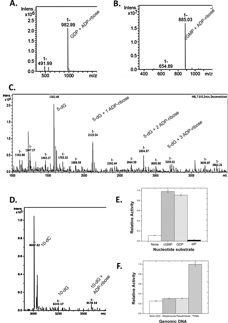
FIGURE 3.
Mass spectrometry of Scabin with oligonucleotide substrates. A, product ion spectra (singly charged, positive mode) after liquid chromatographic separation of the reaction products from the incubation of Scabin with 0.5 mm GDP. B, product ion spectra (singly charged, positive mode) after liquid chromatographic separation of the reaction products from the incubation of Scabin with 0.5 mm cGMP. C, product ion spectra (singly charged, positive mode) after liquid chromatographic separation of the reaction products from the incubation of Scabin with annealed poly(5)-deoxyguanidine/deoxycytidine oligonucleotide. Peaks corresponding to unlabeled (1582.5 Da), singly labeled (2123.5 Da), doubly labeled (2834.4 Da), and triply labeled (3205.6 Da) oligonucleotide were clearly resolved. D, product ion spectra (singly charged, positive mode) after liquid chromatographic separation of the reaction products from the reaction of Scabin with annealed poly(10)-deoxyguanidine/deoxycytidine oligonucleotide. Peaks corresponding to unlabeled (3038.6 Da) and singly labeled (3579.3 Da) oligonucleotide are shown. E, histogram showing the relative activity of Scabin against the following substrates: none (baseline GH activity only), cGMP, GDP, and 2′-deoxyinosine-5′-monophosphate at 1 mm concentrations in GH buffer. F, histogram showing the relative transferase activity of Scabin against genomic DNA from the following organisms: none (GH background activity), S. scabies, P. aeruginosa, and Solanum tuberosum (potato).

