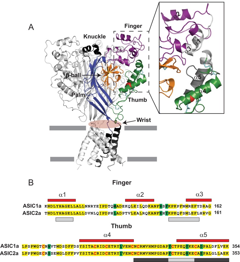
FIGURE 1.
Architecture of the finger and thumb domains. A, schematic of cASIC1 in the desensitized state, illustrating the domain organization of each subunit (PDB code 4NYK). Disulfide bridges are shown as cyan sticks. The Cl− ion bound to the thumb domain is shown in red. mASIC1a and cASIC1 share 89.6% amino acid identity. Inset, close-up view of the finger and thumb domains. The area within the finger and thumb domains examined in this work is shown in gray. Note that the light gray region in the finger domain (Tyr109-Leu114 and Lys148-Tyr158) is in close proximity to the light gray region in the thumb domain (Tyr334-Glu342). The region highlighted in the thumb domain in dark gray is located near the β-ball domain in the desensitized state. B, sequence alignment of the mASIC1a and mASIC2a finger and thumb domains. mASIC1a numbered α helices (red) are shown above their corresponding amino acids. Identical residues are highlighted in yellow, whereas conserved residues are highlighted in green. Conserved cysteine residues are shown in red. The areas within the finger and thumb domains examined in Figs. 5 and 6 are shown in light and dark gray (see above).

