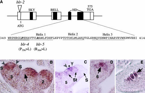
Figure 2.
Molecular Analyses of BLR, a BELL Class Homeodomain Protein.
(A) A diagram illustrating the BLR gene organization and the molecular lesions in blr-2, blr-4, and blr-5. The four exons are indicated by the four boxes connected by a line. The locations of the SKY, BELL, and homedomain (HD) are indicated by closed regions. The amino acid sequence in the homeodomain is shown. The three α helices and the N-terminal arm are underlined by dotted and solid lines, respectively. The amino acids affected by blr-4 and blr-5 are in bold. Numbers indicate the amino acid.
(B) to (E) In situ hybridizations of 8-μm longitudinal sections of inflorescences using a BLR 3′ probe. This probe detected no signal in blr-2 tissues. Numbers indicate the stage of each flower.
(B) In the shoot apex, an arrow indicates the exclusion of BLR mRNA from an emerging FM. In the stage 2 flower, BLR mRNA is present in the peripheral zone. In the stage 3 flower, BLR mRNA becomes excluded from the sepal primordia (S). A narrow band of BLR-expressing cells (marked by a pair of arrowheads) flanks the center of the stage 3 flower.
(C) A stage 5 flower showing a narrow BLR-expressing band (marked by a pair of arrowheads) situated between stamen (St) primordia and the remaining central zone marked by an asterisk.
(D) BLR is localized in the center (arrow) of a stage 6 flower.
(E) BLR mRNA is detected in the chalazal (arrow) domain of developing ovules.

