Figure 5.
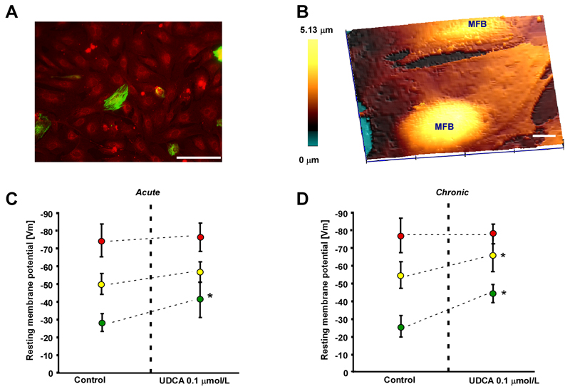
Effect of ursodeoxycholic acid (UDCA) on membrane potential (Vm) in the maternal heart model (MH), fetal heart model (FH) and myofibroblast monolayers. A. Myofibroblast monolayer stained for desmin (green) and vimentin (red) indicates a low contamination by desmin positive cardiomyocytes. Bar=100 μm. B. Scanning ion conductance microscopy identifies the most prominent area for impalement as regions above nuclei in the myofibroblast monolayer. Bar=10 μm. C. Acute exposure to 0.1 μmol/L UDCA significantly increases Vm only in pure myofibroblast monolayers (green dots) but not in the MH model (red dots) and FH model (yellow dots). D. Same as C for chronic exposure to UDCA. 0.1 μmol/L. UDCA significantly increases Vm in the FH model and myofibroblast monolayers but has no effect on MH model preparations. Data: means±SD, n=12.

