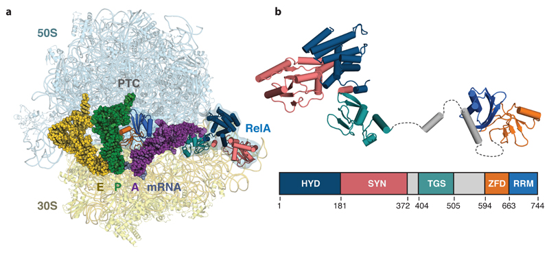
Figure 1. Structure of RelA bound to the ribosome.
a, Overall view of RelA in complex with a ribosome stalled with an uncharged tRNA in the A-site. Displayed are the 50S and 30S ribosomal subunits; E-, P- and A-site tRNAs; mRNA, and RelA coloured by domain. b, Structure of the ribosome-bound form of RelA oriented from N- to C-terminus with the domain organization below showing the boundaries of the hydrolase (HYD), synthetase (SYN), TGS, Zinc-finger (ZFD) and RNA recognition motif (RRM) domains. Unmodeled flexible elements that connect RelA domains are indicated with dashed lines.

