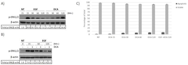
Fig. 1. DCA induced activation of EGFR-MAPK signaling.
HT-29 cells were grown to 80% confluence and then serum starved for 18 hours prior to treatments. (A) Cell cultures were either not treated (NT) or treated with 100 ng/ml EGF or with 250 μM DCA alone for the times indicated. (B) HT-29 cells were either not treated (NT) or treated with 100 ng/ml EGF only or in combination with 250 μM DCA for the times indicated. Lysates were prepared from the treated cells and phosphorylated ERK1/2 was detected by immunoblotting of the SDS-PAGE fractionated proteins. β-actin was used as the loading control. Densitometry values were calculated as described in methods. The results for a typical experiment are shown. (C) Acridine Orange and Ethidium Bromide assay was performed to detect levels of apoptosis in HT-29 cells treated with 250 μM DCA or DCA plus 100 ng/ml EGF at times indicated (min.).

