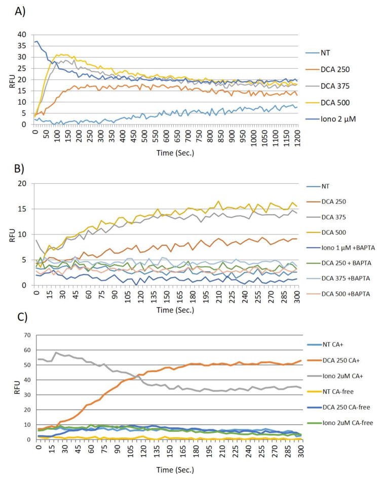
Fig. 3. DCA induces Ca2+ influx.
(A) HT-29 cells were grown in 96 well plates, serum starved as described in Figure 1, and then treated with DCA at the concentrations indicated. Thirty reads per well were taken every 10 seconds for 20 minutes. (B) HT-29 cells were preincubated with 20μM BAPTA-AM for 30 minutes prior to treating with DCA. Sixteen readings per well were taken every 5 seconds for 5 minutes total. C) Calcium assay was performed as previously described (methods), Calcium-free conditions were created by utilizing calcium-free assay buffer in place of traditional assay buffer. Treatment with DCA or ionomycin at the concentrations indicated, were carried out in calcium free conditions (CA-free) where indicated or conventional calcium-containing assay buffer (CA+). Kinetic reads were taken every 10 sec for 20 min. Data points show the average of two wells and all points are normalized to the lowest read per plate. HT-29 cells incubated with DCA and ionomysin at the concentrations indicated were used for comparison. These experiments were performed nine times. The results from a typical experiments are depicted.

