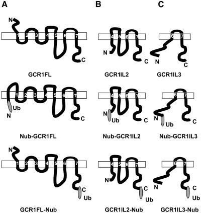
Figure 3.
Topology Prediction of GCR1 Fragments Fused with Ubiquitin at the N or C Terminus as Used for Split Ubiquitin Assays.
Schematic representation of topology of GCR1 full-length and truncated proteins with or without a ubiquitin (N-terminal part, Nub) fusion. Topology was predicted using transmembrane domain hidden Markov model (TMHMM version 2.0). Rectangles represent plasma membrane. Predicted transmembrane regions are numbered from 1 to 7. Ub represents N-terminal portion of ubiquitin.
(A) GCR1 full-length protein (GCR1FL) without or with Ub fusion at the N terminus (Nub-GCR1FL) or at the C terminus (GCR1FL-Nub).
(B) GCR1IL2 protein (amino acids 105 to 326) without or with Ub fusion at the N terminus (Nub-GCR1IL2) or at the C terminus (GCR1IL2-Nub).
(C) GCR1IL3 protein (amino acids 271 to 326) without or with Ub fusion at the N terminus (Nub-GCR1IL3) or at the C terminus (GCR1IL3-Nub).

