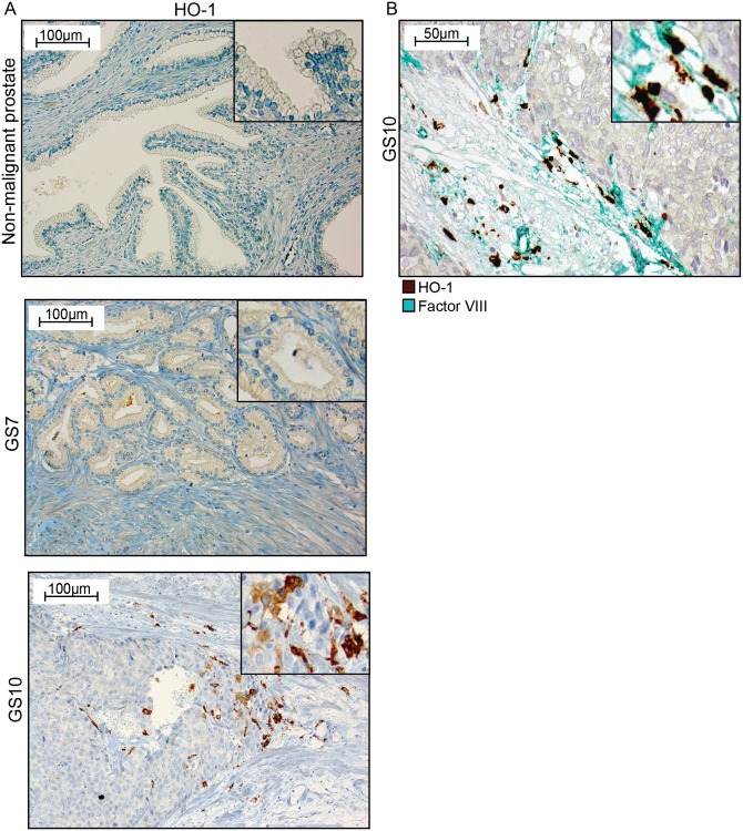
Fig 6. HO-1 expression in human primary prostate tumors.
(A) Representative sections of HO-1 staining (brown) in non-malignant prostate tissue, in a Gleason score (GS) 7 primary prostate tumor, and in a GS 10 primary prostate tumor (200x magnifications, inserts show higher magnifications). (B) A GS 10 tumor double stained for factor VIII positive blood vessels (green) and HO-1+ cells (brown) (400x magnifications, and insert at higher magnifications).

