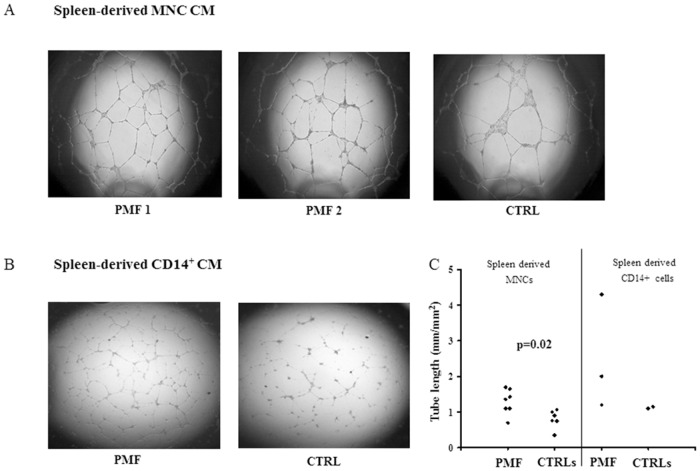
Fig 3. In vitro tubulogenesis induced by spleen MNC or selected CD14+ cell CM.
Digital images of endothelial tubes obtained by bright-field light microscopy 24 hours after plating Endothelial Colony Forming Cells from a healthy donor on Matrigel-coated wells in presence of CM of spleen tissue derived MNCs from two patients with primary myelofibrosis (PMF) and one representative CTRL (panel A), or CM of spleen tissue derived selected CD14+ cells from one representative patient with PMF and one CTRL (panel B). Cultures were examined under an inverted microscope (Labovert, Leitz, Germany) in bright field, at 2.5X magnification using a PL (Leitz Wetzlar, Germany) objective; quantitative evaluation of the tube-like structures, expressed as ratio of the total length of tubular structures per field (mm)/surface of the field (mm2) (ImageJ software by National Institutes of Health, USA, http://rsbweb.nih.gov/ij/.), of all the patients with PMF and CTRLs tested is shown (panel C).

