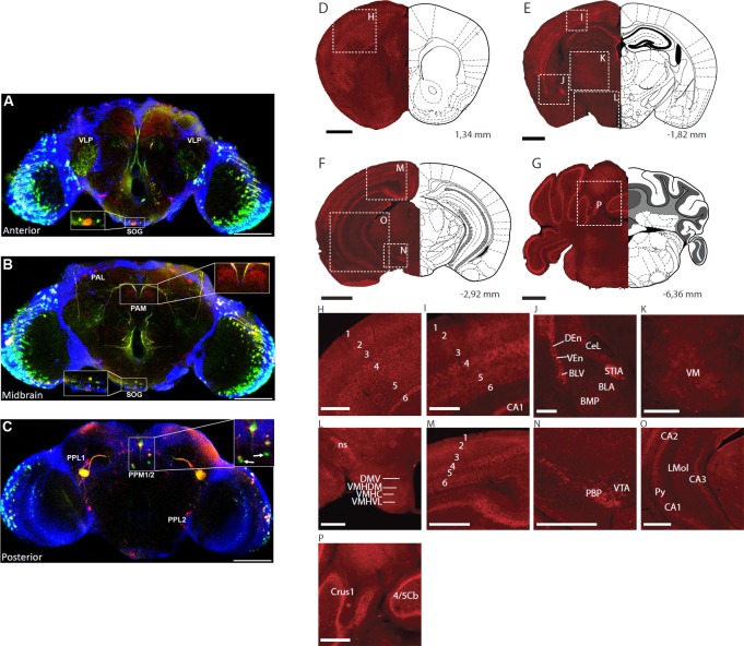
Fig 7. Ets96B and Etv5 expressed on dopaminergic neurons.
(A-C) Ets96B-GAL4 was crossed to UAS-GFP to examine Ets96B expression in adult male brains. Adult male brains were then stained for GFP and Tyrosine hydroxylase (TH) expression. (A) Anterior section showing co-expression of GFP and TH in the eye, the ventrolateral prototcerebrum (VLP and the suboesophageal ganglion (SOG, see inset). (B) Midbrain, no co-expression was observed in the dopaminergic clusters PAM or PAL, while coexpression was observed in the SOG (see inset). (C) In the posterior brain section no co-expression was observed in dopaminergic clusters PPL1 or PPL2, but there was co-expression of GFP and TH in cluster PPM1/2 (see inset). There were also two neurons near the dopaminergic PPM1/2 cluster that were GFP specific, and had no TH expression (see inset, white arrow). In A-C size bar is equivalent to 100 μm. (D-G) Black scale bar, 1mm. (H-P) white scale bar, 0.5 mm. Bregma levels and described brain regions are according to Allen Mouse Brain Atlas. (H, I) Cortex layers 1–6. (J) basolateral amygdaloid nucleus, anterior part (BLA), basolateral amygdaloid nucleus, ventral part (BLV), basomedial amygdaloid nucleus, posterior part (BMP), central nucleus of amygdala, lateral part (CeL), dorsal endopiriform claustrum (DEn), stria medullaris (STIA), ventral endopiriform claustrum (VEn), (K) ventromedial thalamic nucleus (VM), (L) dorsomedial hypothalamic nucleus, ventral part (DMV), nigrostriatal tract (ns), ventromedial hypothalamic nucleus, central part (VMHC), ventromedial hypothalamuc nucleus, dorsomedial part (VMHDM), ventromedial hypothalamic nucleus, ventrolateral part (VMHVL), (M) Cortex layers 1–6, (N) parabrachial pigmented nucleus of the VTA (PBP), ventral tegmental area (VTA), (O) field CA1 of the hippocampus (CA1), field CA2 of the hippocampus (CA2), field CA3 of the hippocampus (CA3), lacunosum moleculare layer of the hippocampus (LMol), pyramidal layer of the hippocampus (Py), (P) crus 1 of the ansiform lobule (Crus1), lobule 4 and 5 of the cerebellar vermis (4/5Cb).

