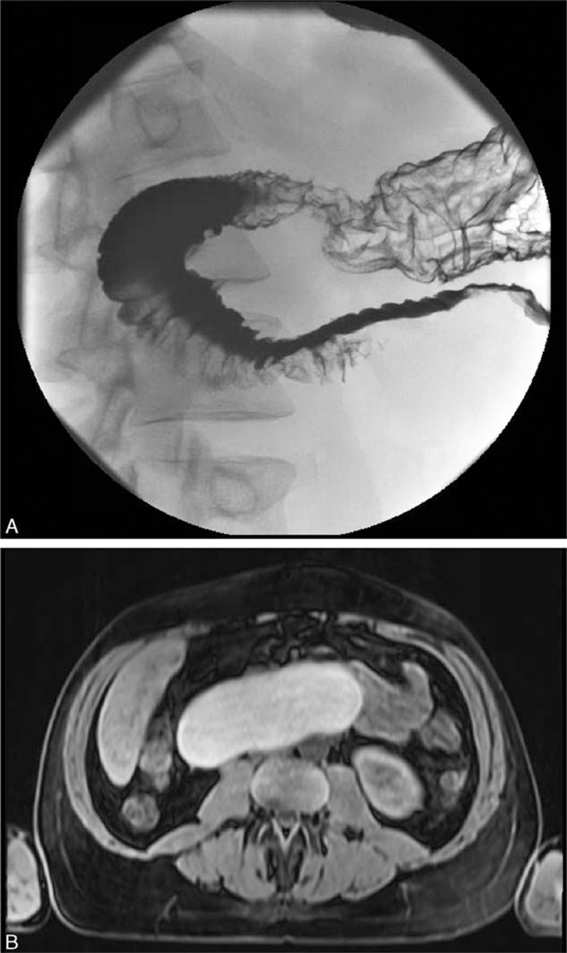
FIGURE 1.

A, Upper gastrointestinal series with small bowel follow-through in a 40-year-old man presenting with severe nausea and vomiting post cibum associated with 15 kg weight loss during the prior month reveals a smoothly contoured deformity along the third portion of the duodenum caused by a central mass displacing and compressing this part of the duodenum. The duodenum is mostly but not completely obstructed by the central mass. B, Abdominal magnetic resonance imaging (MRI) without IV gadolinium contrast reveals an 8 × 13 cm intramural cystic mass arising along wall of the descending and transverse duodenum that demonstrated T1 and T2 hyperintense signals, and an irregularly thickened posterior cyst wall, findings consistent with a duodenal duplication cyst containing complex fluid, such as hemorrhagic or proteinaceous material.
