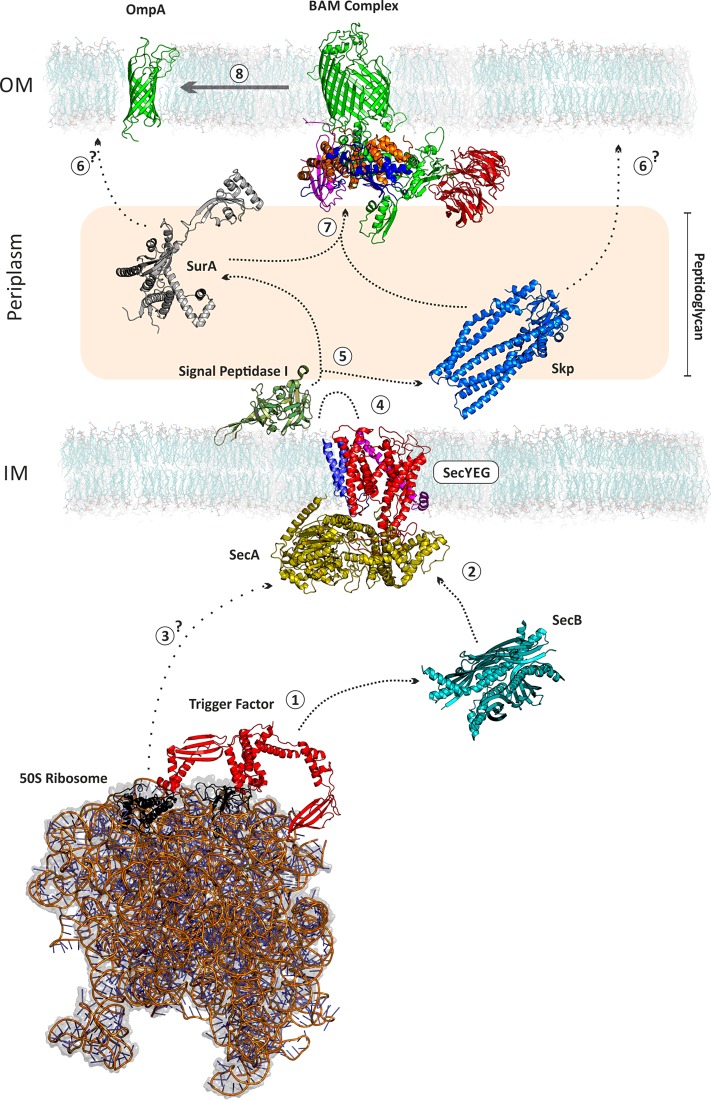
Figure 2. Biogenesis of OMPs.
A nascent OMP emerges from the ribosome and is bound by trigger factor (1) before being passed to SecA via SecB (2), alternatively nascent chains may interact directly with SecA (3). The unfolded OMP (uOMP) passes through the SecYEG channel and the signal sequence is inserted into the inner membrane (IM) (4). This sequence is cleaved by signal peptidase I and the uOMP is bound by the chaperones Skp and/or SurA (5). The uOMP can then be delivered directly to the outer membrane (OM) (6) or to the BAM complex (7). The BAM complex then catalyses the OMP's folding into the OM (8). SecYEG complex: SecY–red, SecE–magenta, SecG–blue, SecA–yellow. BAM complex: BamA–green, BamB–red, BamC–blue, BamD–orange, BamE–magenta. All proteins are shown to scale. The length of the periplasmic space from leaflet to leaflet is scaled to 180 Å. PDB ID of structures: OmpA (1G90); BamACDE (5EKQ); BamB (4XGA); SurA (1M5Y, missing regions built using MODELLER); Skp (1U2M, missing regions built using PyMol); signal peptidase I (1KN9); SecYEG+SecA (3DIN); SecB (1OZB); Trigger Factor (3GU0); 50S ribosome (2D3O). DMPC membrane from O'Neil et al. [21].

