Figure 4.
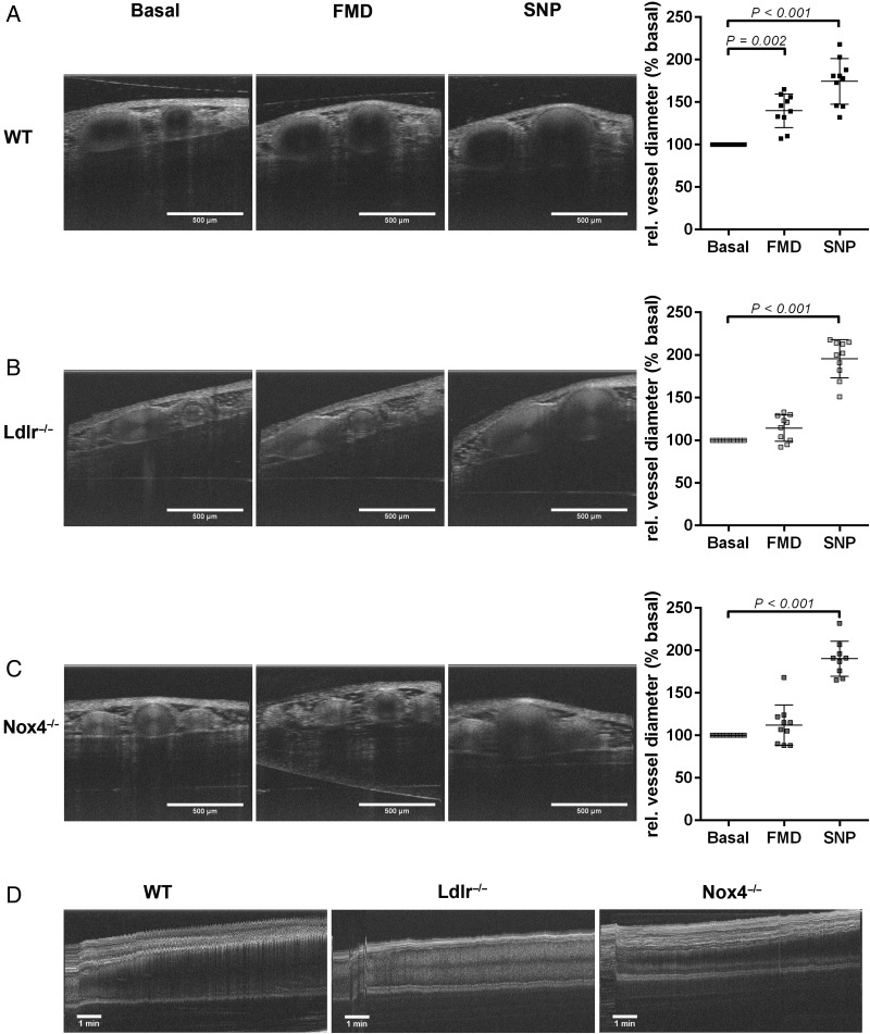
Nox4−/− mice show altered flow-mediated dilation in vivo. (A) Representative optical coherence tomography images of Arteria saphena and evaluation of images of 26-week-old (A) wild-type, (B) Ldlr−/−, and (C) Nox4−/− mice under control conditions (basal), flow-mediated dilation and after application of sodium nitroprusside. (D) Representative optical coherence tomography scan of the Arteria saphena of 26-week-old wild-type, Ldlr−/−, and Nox4−/− mice showing the process of flow-mediated dilation during 10 min after occlusion (n = 10).

