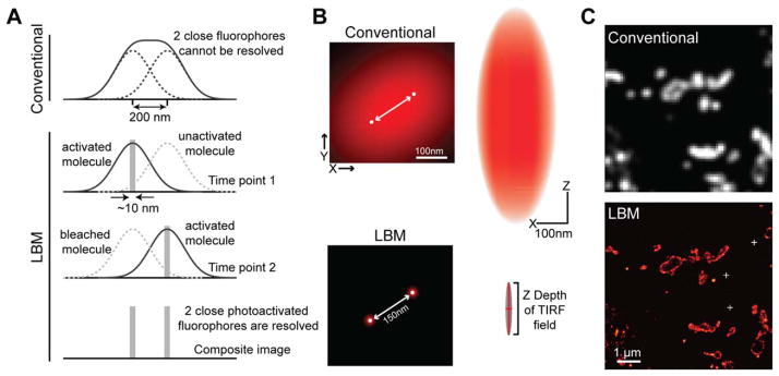
Fig. 1.
Mechanism and demonstration of LBM. A: Under a conventional microscope, two nearby fluorophores cannot be distinguished from each other because their PSFs overlap (top). LBM solves this problem using photoactivatable fluorophores (bottom). Each photoactivatable fluorophore is sequentially activated so that individual molecules can be imaged in isolation and precisely localized. In the composite image where the localization information of all molecules is overlaid, the two fluorophores are resolved [modified from Zhong (2010); with permission]. B: Schematic illustration of the resolving power and the PSF of LBM [modified from Long et al. (2014); with permission]. Left, the effective PSFs of two fluorophores shown in the x–y plane. Right, the PSF is shown in the x–z plane. C: Representative conventional microscopy and LBM images of membrane-tagged EosFPs expressed in fly brain. The sample was processed as described in the Focus Box. White crosses in the LBM image mark the position of gold fiducials for drift correction.

