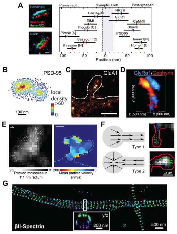
Fig. 2.
Representative studies of LBM in neuroscience. A: The localization of 10 synaptic proteins in thin sections of brain tissue [modified from Dani et al. (2010); with permission]. B: The postsynaptic scaffold protein PSD-95 forms nanometer clusters in cultured hippocampal neurons [modified from MacGillavry et al. (2013); with permission]. C: AMPA receptor GluA1 forms nanoclusters in cultured hippocampal neurons [modified from Nair et al. (2013); with permission]. D: Dual color imaging of the glycine receptor GlyRα1 and the scaffold protein Gephyrin in cultured spinal cord neurons [modified from Specht et al. (2013); with permission]. E: sptPALM tracking the flow of actin at the dendritic spine in cultured hippocampal neurons [modified from Frost et al. (2010); with permission]. Left shows the spatial distribution of actin movement directions in a spine, and right shows the mean actin movement velocity in a different spine. F: sptPALM tracking the flow of GluA1 movement in dendritic spines in hippocampal neurons, identifying two modes of GluA1 dynamics [modified from Hoze et al. (2012); with permission]. Schematics of the results (left) and representative images (right) are shown. G: The actin-associated cytoskeletal protein Spectrin forms a repeated structure in axons of cultured hippocampal neurons [modified from Xu et al. (2012); with permission].

