Figure 1.
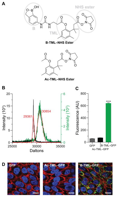
Cellular internalization of B-TML–labeled GFP. (A) Structures of B-TML–NHS ester and Ac-TML–NHS ester. Ellipses denote the three distinct modules within B-TML–NHS ester. (B) MALDI–TOF mass spectra of B-TML–GFP (green), conjugated to ~3 boronic acid moieties per molecule, and the same protein after exposure to CHO K1 cell lysate and purification (gray). Expected m/z: GFP, 29361; each B-TML moiety, 450. (C) Flow cytometry analysis of CHO K1 cells incubated with 10 μM unlabeled GFP, GFP labeled with a control vehicle (Ac-TML), or GFP labeled with the boronate vehicle (B-TML) for 4 h (p < 0.0001). (D) Confocal microscopy of CHO K1 cells grown as in panel C. Cells were stained with WGA-594 (red) and Hoechst 33342 (blue). Scale bars: 10 μm.

