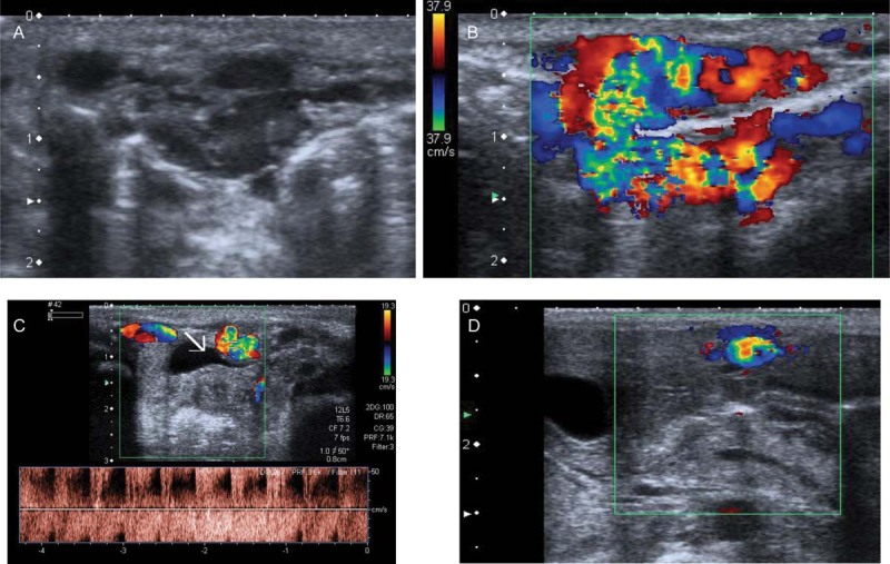
Fig. 1.

(A) Abdominal US and (B) Doppler US: 28 × 16 mm cluster, with a high density of vascularization, mixed arterial and venous. (C) Multiple feeding arteries coming from the external iliac, hypogastric, epigastric, and mammary arteries. (D) Enlarged umbilical vein, venous drainage of the lesion. US, ultrasound.
