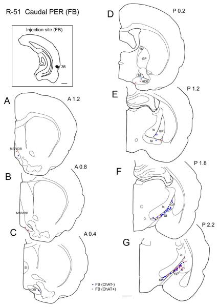
Figure 3.
Distribution of cholinergic and non-cholinergic retrogradely labeled cells following an injection of FB into the caudal portion of the perirhinal cortex (case R-51 FB). Conventions as in Figure 1. Note that, as in Figures 1 and 2, labeling is present densely in the caudal BF (caudal GP and SI) (F, G) and decreases rostrally. Cholinergic projection neurons are present densely in the caudal part of caudal GP (G). Scale bar = 1 mm.

