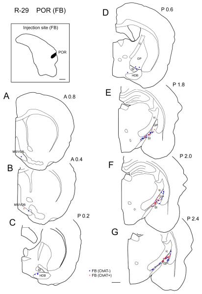
Figure 4.
Distribution of cholinergic and non-cholinergic retrogradely labeled cells following an injection of FB into the postrhinal cortex (case R-29 FB). Conventions as in Figure 1. Note that, as in perirhinal cases (Figs. 1-3), labeling is dense in the caudal BF (caudal GP and SI) (E-G) and decreases rostrally (A-D). Cholinergic projection neurons are labeled densely in the caudal part of caudal GP (F, G). Scale bar = 1 mm.

